Unit 3
Cells
Use a microscope to meet the tiny building blocks of every living thing, and compare plant cells with animal cells.
Read
The building blocks of life
In 1665 an English scientist named Robert Hooke sliced a thin piece of cork, put it under a microscope he had built, and saw rows of tiny box-shaped spaces. They reminded him of the small rooms, called cells, where monks lived. He had given a name to something that every living thing is made of, though he had no idea yet how important it was. Ten years later, in the Netherlands, Antonie van Leeuwenhoek turned a tiny glass lens on pond water and found a whole hidden world of moving creatures, each one a single cell.
The cell theory
After two centuries of looking, scientists agreed on three ideas, now called the cell theory:
- All living things are made of one or more cells.
- The cell is the basic unit of structure and function of life: it is the smallest thing that can carry out the processes of life by itself.
- New cells come only from cells that already exist (by cell division).
Some organisms are a single cell: an Amoeba, a Paramecium in a pond, a yeast cell, a bacterium. They are unicellular. Others, such as a banyan tree or a person, are multicellular. An adult human is built from tens of trillions of cells, of many different kinds: nerve cells that can be a metre long, flat skin cells, round red blood cells that carry oxygen, and muscle cells that can shorten.
Misconception. "Plant cells are square and animal cells are round." Cell shapes depend on the job. Nerve cells are long and branched, red blood cells are discs, and some plant cells are long tubes that carry water. What really separates plant from animal cells is which parts they have, not their outline.
Parts of a cell
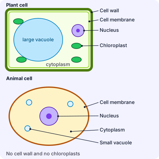
Part What it does Plant Animal Cell membrane A thin, flexible skin that controls what goes in and out yes yes Cytoplasm The jelly-like fluid where many of the cell's chemical reactions happen yes yes Nucleus Holds the cell's instructions (its DNA) and directs the cell's activities yes yes Cell wall A stiff outer layer that supports the cell and gives it a fixed shape yes no Chloroplasts Green structures that trap light to make food (photosynthesis) yes no Vacuole A sac of fluid; in a mature plant cell it is one large vacuole that helps keep the cell firm large small, if any There are other parts too. Mitochondria release energy from food, but they are too small to see with a school microscope. In the next unit you will see why that matters.
Bacteria are cells too, but a simpler kind: they have no nucleus, and their instructions float in the cytoplasm.
Using a light microscope
A cell is usually between about 0.01 mm and 0.1 mm across. The unit used for such tiny lengths is the micrometre (), a thousandth of a millimetre, so a cell might be 10 to 100 m. Your unaided eye can barely see something of 0.1 mm, so we need a microscope to magnify the image.
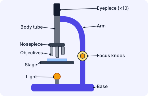
The total magnification is the eyepiece magnification multiplied by the objective magnification:
Example. An eyepiece of with an objective of gives , written .
Good habits at the microscope:
- Start with the lowest-power objective. It has the biggest field of view, so it is easy to find your specimen.
- Use the coarse focus only with low power. With higher power, use only the fine focus, or you can crash the lens into the slide.
- Light comes from a built-in lamp or a mirror. Never point a mirror at the Sun: it can permanently damage your eyes.
- Move to a higher power only once the image is sharp, and re-focus each time.
What you will see
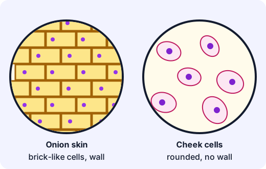
An onion skin peel (a thin, clear layer from the inside of a bulb scale) is the classic plant slide. A drop of iodine or red ink stain makes the nuclei show up. You see tidy, brick-like cells with a clear boundary, the cell wall, and one nucleus each. You will not see chloroplasts because the underground bulb does not make food from light.
Cheek cells (scraped gently from the inside of the cheek by a trained teacher or an adult, with fresh clean tools and a stain such as methylene blue) look rounded and irregular, with a nucleus but no cell wall. For safety this is done only with supervision, each person uses their own clean stick, and everything is washed or thrown away afterwards.
Finding the real size
A microscope makes the picture bigger, so what you draw is not the real size. To find the real size:
Example. You draw an onion cell that is 25 mm long in your drawing, seen at . Its real length is , which is (since ).
Example. A cheek cell is across. At it would appear across. That is easy to draw in your notebook.
Why "cell" is such a powerful idea
Cells tell us what life is made of. A tissue is a group of similar cells working together (muscle tissue), an organ is a team of tissues (the heart), and an organism is a team of organs. A single cell has everything it needs for life, so to understand a living thing, you can start by understanding its cells. In the next unit we ask what those cells are doing when something is alive.
Key ideas
- All living things are made of cells; the cell is the unit of life; new cells come from old cells.
- Plant and animal cells both have a membrane, cytoplasm and a nucleus. Plant cells also have a cell wall, chloroplasts and a large vacuole.
- Total magnification = eyepiece objective; real size = image size magnification.
- Always start at low power, use fine focus at high power, and never use sunlight with a mirror.
Lab
Virtual microscope
Use a virtual light microscope. Choose a slide and an objective, focus until the image is sharp, then compare onion skin (plant cells) with cheek cells (animal cells).
Your goals
- Focus the onion skin sharply at total x100 and x400, and the cheek cells at x400.
- Answer three questions about the nucleus, the cell wall and refocusing.
The interactive lab opens once you register. Register to do this.
Quick check
Cells check
Eight questions on cells and microscopes.
1.What is the main job of the nucleus?
- It holds the cell's instructions and directs its activities.
- It traps light to make food.
- It gives the cell a stiff outer layer.
- It lets water leave the cell.
Choose one
2.Which parts are found in a plant cell but NOT in an animal cell? Pick all that apply.
- Cell wall
- Nucleus
- Chloroplasts
- Cell membrane
- Large central vacuole
Choose all that apply
3.A microscope has a x10 eyepiece and you are using the x40 objective. What is the total magnification?
Answer with a number
4.A cell looks 6 mm wide in an image magnified x400. What is its real width, in millimetres?
Answer with a number
5.A slide is already on the stage. Put these steps in the safest order.
- Click the lowest-power objective into place
- Sharpen the image with the fine focus
- Use the coarse focus to bring the cells into view
- Switch to a higher-power objective and sharpen with the fine focus only
Put these in order
6.An Amoeba in pond water moves, feeds and divides, yet under the microscope it is just one cell. What does this tell us?
- One cell can be a whole living organism.
- It cannot be alive because it is so small.
- It must be a plant cell.
- Only animals can be a single cell.
Choose one
7.Match each cell part to its job.
- Cell membrane
- Cytoplasm
- Chloroplast
- Cell wall
Match each to: Traps light energy to make food · Stiff layer that supports a plant cell · Jelly where many chemical reactions happen · Controls what enters and leaves the cell
8.Which statement about cell shapes is correct?
- All plant cells are square and all animal cells are round.
- Cells come in many shapes, each suited to its job.
- All cells in one body look the same.
- A cell's shape never matters.
Choose one
Answering and checking are for registered learners. Register to do this.
Quick check
New puzzles on cells
Use what you know about cells in situations the reading did not show.
1.A human hair looks 8 mm wide in an image magnified x100. What is the real width of the hair, in millimetres?
Answer with a number
2.Each cell in a thin layer is 25 micrometres (25 μm) across. How many such cells placed side by side in a row would stretch 1 mm? (1 mm = 1000 μm.)
Answer with a number
3.A student says her slide is a thin layer from a green leaf. Under the microscope she sees rounded cells with a nucleus and a membrane, but no cell wall and no chloroplasts. What is the best conclusion?
- The slide is probably from an animal, because leaf cells have a cell wall and chloroplasts.
- The slide is from a plant, because all cells have a nucleus.
- Leaves have no cells.
- The microscope must be broken, because plant cells can have no wall.
Choose one
4.A scientist puts a drop of bubbly yeast mixture under the microscope and sees many separate oval cells, each with its own nucleus, some with a small bud growing off them. What does this show?
- Yeast is made of one cell, and each cell can make new cells by budding.
- Yeast is not alive because it has no cell wall.
- Yeast is a multicellular animal.
- Yeast has no nucleus.
Choose one
Answering and checking are for registered learners. Register to do this.
Your turn
Explain cells
Write in your own words. A simple sketch may help (describe it in words if you cannot upload one).
- In your own words, explain the cell theory and why a single Amoeba is as much "alive" as an elephant.
- Compare a plant cell and an animal cell. Name two parts both have and two parts only the plant cell has, and say what each part does.
- A drawing of a cell is 36 mm long. The microscope view was at x400. Show how you work out the real length, in millimetres and in micrometres.
- Describe the steps you would follow to get a sharp image on a microscope, and give one safety rule.
How it's judged
- Cell theory
- Comparing plant and animal cells
- Calculating real size
- Using a microscope
Writing and submitting your answer is for registered learners. Register to do this.
Like what you see?
Register for free to answer the quick checks, do the activities and save your work. Your work is saved on your device and syncs to your account.